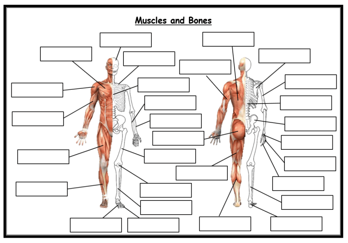
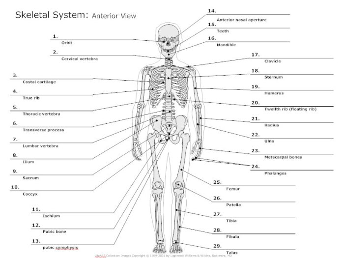
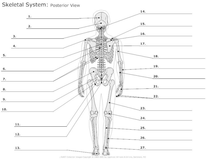
Fill in the blanks in the skeletal diagram. – Embark on an enlightening journey into the realm of skeletal diagrams, where the meticulous placement of bones, joints, and muscles unveils a wealth of anatomical insights. Dive into the intricacies of constructing and interpreting these diagrams, unlocking the secrets of human movement, posture, and pathology.
As we delve deeper into the topic, we will explore the diverse applications of skeletal diagrams, from their indispensable role in medical diagnosis to their captivating use in education and research. With each step, we unravel the techniques and strategies that empower you to confidently fill in the blanks, completing the skeletal puzzle and revealing the wonders of human anatomy.
1. Skeletal Diagram Overview

A skeletal diagram is a schematic representation of the skeletal system, depicting the bones, joints, and muscles in a simplified and organized manner. Its primary purpose is to provide a visual representation of the human skeletal structure, aiding in the understanding of anatomy, biomechanics, and related fields.
Skeletal diagrams are invaluable in various fields, including:
- Medicine:Assisting in the diagnosis and treatment of skeletal disorders, surgical planning, and patient education.
- Education:Enhancing the comprehension of human anatomy in medical schools, biology classes, and fitness programs.
- Research:Facilitating the study of skeletal development, biomechanics, and evolutionary biology.
- Forensic science:Aiding in the identification and analysis of skeletal remains.
2. Types of Skeletal Diagrams
There are various types of skeletal diagrams, each tailored to specific purposes:
- Anatomical Diagrams:Provide a comprehensive view of the skeletal system, including all bones, joints, and muscles.
- Biomechanical Diagrams:Focus on the mechanical properties of the skeletal system, depicting forces, moments, and stresses acting on bones and joints.
- Developmental Diagrams:Illustrate the skeletal development process from infancy to adulthood.
- Pathological Diagrams:Depict skeletal abnormalities, such as fractures, dislocations, and deformities.
- Comparative Diagrams:Compare skeletal structures of different species, highlighting evolutionary relationships and adaptations.
3. Elements of a Skeletal Diagram

Essential elements of a skeletal diagram include:
- Bones:Represented as lines or shapes, labeled with their anatomical names.
- Joints:Indicated by circles or other symbols, denoting the points of articulation between bones.
- Muscles:Attached to bones and depicted as shaded areas or lines, illustrating their origin and insertion points.
- Ligaments and Tendons:May be shown as lines or dots, connecting bones and muscles, respectively.
- Labels and Annotations:Provide detailed information about the skeletal structures, such as bone names, joint types, and muscle functions.
4. Constructing a Skeletal Diagram: Fill In The Blanks In The Skeletal Diagram.

Constructing a skeletal diagram involves:
- Gather Information:Collect anatomical data from textbooks, atlases, or online resources.
- Sketch the Artikel:Draw a basic Artikel of the skeletal structure, including the major bones and joints.
- Add Details:Gradually add bones, joints, muscles, and other elements based on the information gathered.
- Label and Annotate:Provide clear labels and annotations to identify the structures accurately.
- Review and Refine:Check for accuracy, completeness, and clarity. Make necessary adjustments to ensure the diagram is informative and visually appealing.
5. Techniques for Filling in Blanks
To effectively fill in blanks in a skeletal diagram:
- Identify Missing Information:Determine the missing elements based on the context of the diagram and your anatomical knowledge.
- Consult Resources:Refer to textbooks, atlases, or online databases to gather the necessary information.
- Make Informed Guesses:If specific information is unavailable, make reasonable guesses based on the surrounding structures and anatomical principles.
- Check for Consistency:Ensure that the filled-in elements are consistent with the overall structure and anatomical accuracy.
- Seek Expert Advice:If necessary, consult with an anatomist or medical professional for guidance.
6. Examples and Applications
Skeletal diagrams are widely used in:
- Medical Textbooks and Atlases:Illustrating skeletal anatomy and pathology for students and practitioners.
- Educational Materials:Enhancing the understanding of human anatomy in schools, universities, and fitness programs.
- Research Publications:Depicting skeletal structures and biomechanical data for scientific studies.
- Forensic Investigations:Assisting in the identification and analysis of skeletal remains.
- Patient Education:Providing visual explanations of skeletal conditions and treatment options.
Commonly Asked Questions
What is the primary purpose of a skeletal diagram?
Skeletal diagrams provide a visual representation of the human skeletal system, aiding in the study of anatomy, diagnosis of medical conditions, and design of surgical procedures.
How can I effectively fill in the blanks in a skeletal diagram?
Begin by identifying the known elements and their relationships. Use reference materials, such as textbooks or online resources, to determine the missing information. Make informed guesses based on anatomical knowledge and logical reasoning.
What are the common applications of skeletal diagrams?
Skeletal diagrams are widely used in medical education, patient diagnosis, forensic investigations, and biomechanical research. They provide valuable insights into human movement, posture, and skeletal abnormalities.